Scanning Electron Microscopy (SEM)
Scanning Electron Microscopy (SEM) is a high-resolution imaging technique that uses a focused beam of electrons to scan the surface of a sample, generating detailed topographical and compositional information. When the electron beam interacts with the sample, it produces various signals, including secondary electrons (SE) for surface morphology and backscattered electrons (BSE) for elemental contrast. These signals are detected and processed to form high-magnification images with nanometer-scale resolution. Unlike optical microscopy, SEM provides exceptional depth of field and spatial resolution (down to ~1 nm), making it indispensable for analyzing the morphology, size, and surface characteristics of pharmaceutical particles, nanomaterials, and drug delivery systems.
Applications in Pharmaceutical Partical Analysis
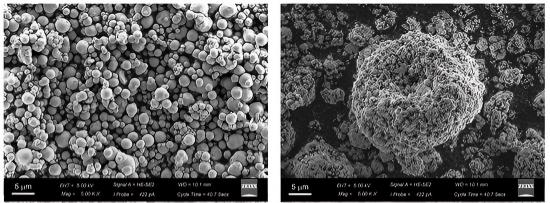
SEM plays a critical role in drug development by enabling direct visualization and characterization of drug formulations, excipients, and particulate contaminants. In solid dosage forms (tablets, capsules), SEM helps assess particle shape, surface roughness, and coating uniformity, which influence dissolution rates and bioavailability. For nanoparticle-based therapeutics (e.g., liposomes, polymeric nanoparticles), SEM provides insights into aggregation, structural integrity, and batch-to-batch consistency. Additionally, SEM is used in failure analysis to identify particulate impurities or defects in injectable drugs, ensuring compliance with FDA and EMA regulatory standards. Its ability to combine imaging with Energy-Dispersive X-ray Spectroscopy (EDS) further allows elemental analysis, crucial for detecting metal contaminants or verifying coating compositions.
SEM offers unparalleled advantages for pharmaceutical particle analysis. Its nanometer-scale resolution surpasses optical microscopy, enabling the detection of sub-micron particles and surface defects that could impact drug performance. The high depth of field allows for clear imaging of rough or porous samples, such as freeze-dried formulations or fibrous excipients. Unlike techniques like Dynamic Light Scattering (DLS) or Nanoparticle Tracking Analysis (NTA), SEM provides direct visual confirmation of particle morphology, eliminating ambiguities in size distribution data. Advanced SEM systems equipped with cryogenic capabilities (Cryo-SEM) can analyze hydrated or temperature-sensitive samples (e.g., protein aggregates, emulsions) without drying artifacts. Furthermore, EDS integration enables simultaneous chemical mapping, identifying elemental composition without requiring separate tests. These capabilities make SEM a gold standard for formulation optimization, quality control, and regulatory submissions.
PharmaAnalytica's Technology Platform
KYKY-EM8200
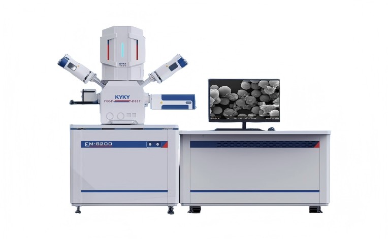
KYKY-EM8000 field-emission SEM provides high-resolution (0.8 nm) imaging for precise analysis of drug particle morphology, coating integrity, and nanocarrier characterization in pharmaceutical development.
SS-150 SEM
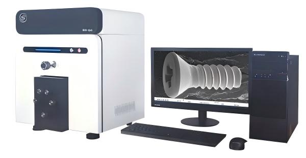
SS-150 benchtop SEM, equipped with dual SE/BSE detectors, enables rapid and clear imaging of drug particle morphology, surface texture, and elemental contrast for efficient pharmaceutical quality control.
PharmaAnalytica's SEM-Based Particle Analysis
PharmaAnalytica's SEM services empower pharmaceutical developers with ultra-high-resolution imaging and elemental analysis, critical for formulation design, stability testing, and troubleshooting. Whether characterizing nanoparticles, tablets, or biologics, our expertise ensures robust data to accelerate drug development.
Expert Sample Preparation & Imaging
We provide specialized sample preparation, including sputter coating, critical point drying, and cryo-fixation, to prevent artifacts and enhance image quality. Our team optimizes imaging parameters (e.g., voltage, working distance) for each sample type.
Multi-Technique Correlation Analysis
STEMart integrates SEM with complementary techniques such as Energy-Dispersive X-ray Spectroscopy (EDS), Focused Ion Beam (FIB), and Atomic Force Microscopy (AFM) to provide a holistic understanding of samples.
Customized Analytical Workflows
We tailor SEM protocols to address specific drug development challenges: low-dose imaging for beam-sensitive biologics, automated particle counting.
Industry-Specific Expertise
Our team includes pharmaceutical scientists and regulatory specialists who understand industry demands, such as pre-formulation screening, GMP-compliant documentation.
Online Inquiry
This site is protected by reCAPTCHA and the Google Privacy Policy and Terms of Service apply.
Need More Information or Request A Quotation?
Related Links