Transmission Electron Microscopy (TEM)
Transmission Electron Microscopy (TEM) is a high-resolution imaging technique that uses a beam of electrons transmitted through an ultra-thin sample to generate detailed structural and compositional information at the atomic or nanoscale level. In TEM, electrons are accelerated under high voltage (typically 80–300 kV) and focused by electromagnetic lenses to interact with the sample. As electrons pass through the specimen, they are scattered or absorbed depending on the material's density and thickness, producing contrast in the resulting image. Advanced detectors capture these interactions, enabling visualization of crystal structures, particle morphology, and even elemental distribution via techniques like energy-dispersive X-ray spectroscopy (EDS) or electron energy loss spectroscopy (EELS). Unlike optical microscopy, TEM achieves resolutions below 0.1 nm, making it indispensable for studying nanoparticles, proteins, and other ultrafine drug components.
Applications in Pharmaceutical Partical Analysis
In pharmaceutical research, TEM provides critical insights into drug formulation and quality control. It enables direct observation of nanoparticle size, shape, and dispersion, essential for optimizing drug delivery systems (e.g., liposomes, polymeric nanoparticles). TEM also characterizes crystallinity and defects in active pharmaceutical ingredients (APIs), which influence solubility and stability. For biologics, TEM visualizes protein aggregates, virus-like particles (VLPs), and antibody-drug conjugates (ADCs), ensuring their structural integrity. Additionally, TEM's ability to map elemental composition (via EDS) helps identify contaminants or coating uniformity in complex formulations. By offering atomic-level details, TEM supports regulatory submissions, accelerates formulation troubleshooting, and enhances understanding of drug behavior in vivo.
TEM surpasses other microscopy techniques in resolution and versatility. Its sub-nanometer resolution reveals ultrafine details, such as lattice fringes in crystals or lipid bilayers in nanocarriers, which are invisible to AFM or SEM. TEM also combines imaging with chemical analysis (EDS/EELS), eliminating the need for separate instruments. Unlike surface-limited techniques (e.g., SEM), TEM provides internal structural information, such as hollow cores in nanoparticles or layered drug-polymer matrices. Cryo-TEM further extends capabilities by preserving hydrated samples (e.g., emulsions, proteins) in near-native states, avoiding artifacts from drying or staining. Moreover, TEM accommodates diverse samples—from inorganic nanoparticles to soft biologics—making it a universal tool for drug development stages, from discovery to manufacturing QC.
PharmaAnalytica's Technology Platform
TH-F120 Field-Emission TEM
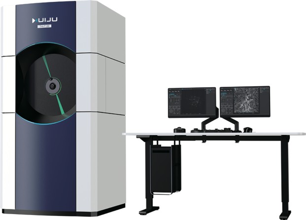
TH-F120 offers information resolution down to 0.14 nm and magnification up to 1,500,000×. It delivers ultra-high resolution for internal structural analysis of liposomes, nanoparticles, nanocrystals, microcapsules and protein assemblies, supporting design and optimization of targeted delivery systems and biologic characterization.
BZ-F200 Field-Emission TEM
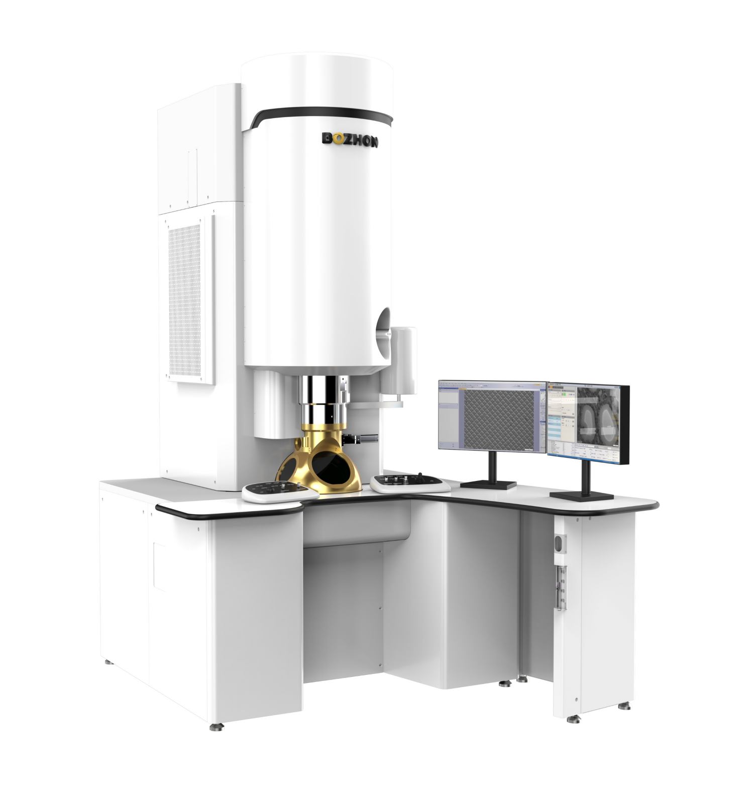
BZ-F200 is a 200kV domestic field-emission TEM with independent IP, optimized for pharmaceutical particle analysis. It has 0.14 nm lattice resolution, 100×-1,200,000× magnification, reduces electron beam damage, enables high-throughput testing, and meets pharmacopoeial standards.
PharmaAnalytica's TEM-Based Particle Analysis
PharmaAnalytica's TEM services empower pharmaceutical teams with unmatched resolution, multimodal imaging, and cryogenic preservation, addressing challenges in nanoparticle characterization, biologics development, and QC.
Ultra-High Resolution Imaging
Our TEM systems achieve sub-nanometer (≤0.1 nm) resolution, enabling visualization of fine structural details. This level of detail is critical for understanding particle morphology, defects, and phase distribution, which directly impact drug performance.
Expert TEM Technical Team
PharmaAnalytica's TEM services are supported by a highly specialized team of electron microscopy scientists which consists of PhD-level experts in nanoparticle imaging, certified TEM operators trained in advanced techniques and regulatory compliance specialists.
Optimized Experimental Design
We tailor TEM experiments to maximize data relevance for pharmaceutical applications. Our experimental designs focus on reproducibility, regulatory alignment, and problem-solving focus.
Regulatory Compliant
Our TEM services adhere to Good Laboratory Practice (GLP) standards, ensuring data reliability for regulatory submissions (FDA/EMA). We provide detailed reports with statistical analysis of particle size, crystallinity, and morphology.
By integrating cutting-edge TEM with regulatory-compliant analytics, we accelerate drug development timelines while ensuring data reliability. Partner with us to transform nanoscale insights into actionable formulation strategies.
Online Inquiry
This site is protected by reCAPTCHA and the Google Privacy Policy and Terms of Service apply.
Need More Information or Request A Quotation?
Related Links